Cell size
Prokaryotic and eukaryotic cells
Prokaryotic cells
Introduction
Take a moment and look at yourself. How many organisms do you see? Your first thought might be that there's just one: yourself. However, if you were to look closer, at the surface of your skin or inside your digestive tract, you would see that there are actually many organisms living there. That’s right - you are home to around 100 trillion bacterial cells!
This means that your body is actually an ecosystem. It also means that you—for some definition of the word you—actually consist of both of the major types of cells: prokaryotic and eukaryotic.
All cells fall into one of these two broad categories. Only the single-celled organisms of the domains Bacteria and Archaea are classified as prokaryotes—pro means before and kary means nucleus. Animals, plants, fungi, and protists are all eukaryotes—eu means true—and are made up of eukaryotic cells. Often, though—as in the case of we humans—there are some prokaryotic friends hanging around.

Components of prokaryotic cells
There are some key ingredients that a cell needs in order to be a cell, regardless of whether it is prokaryotic or eukaryotic. All cells share four key components:
The plasma membrane is an outer covering that separates the cell’s interior from its surrounding environment.
Cytoplasm consists of the jelly-like cytosol inside the cell, plus the cellular structures suspended in it. In eukaryotes, cytoplasm specifically means the region outside the nucleus but inside the plasma membrane.
DNA is the genetic material of the cell.
Ribosomes are molecular machines that synthesize proteins.
Despite these similarities, prokaryotes and eukaryotes differ in a number of important ways. A prokaryote is a simple, single-celled organism that lacks a nucleus and membrane-bound organelles. We’ll talk more about the nucleus and organelles in the next article on eukaryotic cells, but the main thing to keep in mind for now is that prokaryotic cells are not divided up on the inside by membrane walls, but consist instead of a single open space.
The majority of prokaryotic \[\text{DNA}\] is found in a central region of the cell called the nucleoid, and it typically consists of a single large loop called a circular chromosome. The nucleoid and some other frequently seen features of prokaryotes are shown in the diagram below of a cut-away of a rod-shaped bacterium.
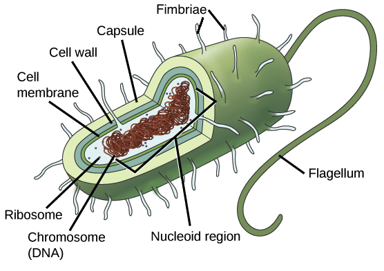
_Image credit: modified from "Prokaryotic cells: Figure 1" by OpenStax College, Biology, CC BY 3.0_
Bacteria are very diverse in form, so not every type of bacterium will have all of the features shown in the diagram.
Most bacteria are, however, surrounded by a rigid cell wall made out of peptidoglycan, a polymer composed of linked carbohydrates and small proteins. The cell wall provides an extra layer of protection, helps the cell maintain its shape, and prevents dehydration. Many bacteria also have an outermost layer of carbohydrates called the capsule. The capsule is sticky and helps the cell attach to surfaces in its environment.
Some bacteria also have specialized structures found on the cell surface, which may help them move, stick to surfaces, or even exchange genetic material with other bacteria. For instance, flagella are whip-like structures that act as rotary motors to help bacteria move.
Fimbriae are numerous, hair-like structures that are used for attachment to host cells and other surfaces. Bacteria may also have rod-like structures known as pili, which come in different varieties. For instance, some types of pili allow a bacterium to transfer \[\text{DNA}\] molecules to other bacteria, while others are involved in bacterial locomotion—helping the bacterium move.
Archaea may also have most of these cell surface features, but their versions of a particular feature are typically different from those of bacteria. For instance, although archaea also have a cell wall, it's not made out of peptidoglycan—although it does contain carbohydrates and proteins.
Cell size
Typical prokaryotic cells range from 0.1 to 5.0 micrometers (μm) in diameter and are significantly smaller than eukaryotic cells, which usually have diameters ranging from 10 to 100 μm.
The figure below shows the sizes of prokaryotic, bacterial, and eukaryotic, plant and animal, cells as well as other molecules and organisms on a logarithmic scale. Each unit of increase in a logarithmic scale represents a 10-fold increase in the quantity being measured, so these are big size differences we’re talking about!
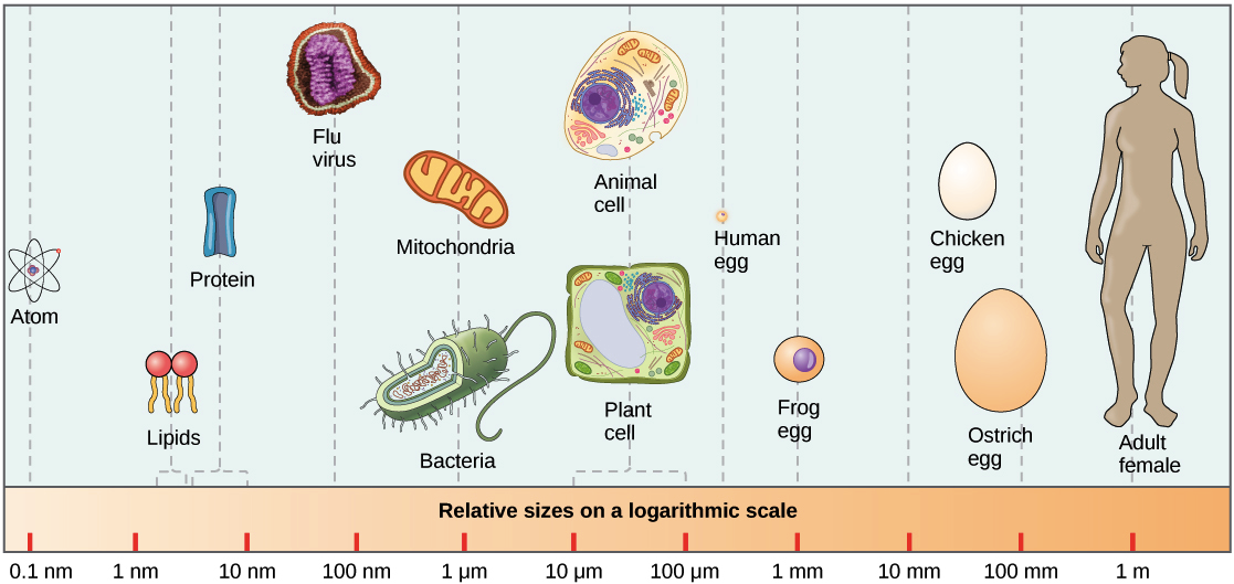
_Image credit: "Prokaryotic cells: FIgure 2" by OpenStax College, Biology, CC BY 3.0_
With a few cool exceptions—check out the single-celled seaweed Caulerpa—cells must remain fairly small, regardless of whether they’re prokaryotic or eukaryotic. Why should this be the case? The basic answer is that as cells become larger, it gets harder for them to exchange enough nutrients and wastes with their environment. To see how this works, let’s look at a cell’s surface-area-to-volume ratio.
Suppose, for the sake of keeping things simple, that we have a cell that’s shaped like a cube. Some plant cells are, in fact, cube-shaped. If the length of one of the cube’s sides is \[l\], the surface area of the cube will be \[6l^2\], and the volume of the cube will be \[l^3\]. This means that as \[l\] gets bigger, the surface area will increase quickly since it changes with the square of \[l\]. The volume, however, will increase even faster since it changes with the cube of \[l\].
Thus, as a cell gets bigger, its surface-area-to-volume ratio drops. For example, the cube-shaped cell on the left has a volume of 1 mm\[^3\] and a surface area of 6 mm\[^2\] with a surface-area-to-volume ratio of six to one, whereas the cube-shaped cell on the right has a volume of 8 mm\[^3\] and a surface area of 24 mm\[^2\] with a surface area-to-volume ratio of three to one.
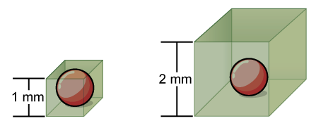
_Image credit: modified from "Prokaryotic cells: FIgure 3" by OpenStax College, Biology, CC BY 3.0_
Surface-area-to-volume ratio is important because the plasma membrane is the cell’s interface with the environment. If the cell needs to take up nutrients, it must do so across the membrane, and if it needs to eliminate wastes, the membrane is again its only route.
Each patch of membrane can exchange only so much of a given substance in a given period of time – for instance, because it contains a limited number of channels. If the cell grows too large, its membrane will not have enough exchange capacity (surface area, square function) to support the rate of exchange required for its increased metabolic activity (volume, cube function).
The surface-area-to-volume problem is just one of a related set of difficulties posed by large cell size. As cells get larger, it also takes longer to transport materials inside of them. These considerations place a general upper limit on cell size, with eukaryotic cells being able to exceed prokaryotic cells thanks to their structural and metabolic features—which we’ll explore in the next section.
Some cells also use geometric tricks to get around the surface-area-to-volume problem. For instance, some cells are long and thin or have many protrusions from their surface, features that increase surface area relative to volume\[^2\].
Intro to eukaryotic cells
Introduction
What would it be like to live in a one-room cabin? Well, things would probably be pretty simple. You would eat, sleep, work, and relax in a single room—which might be a bit cramped, but would certainly make cleaning the house a snap!
Prokaryotic cells, the simple cells of organisms like bacteria, are sometimes compared to one-room cabins: they don't have internal membranes, so they’re like a single room with no walls to carve it up\[^1\]. If we extend this analogy to eukaryotic cells, the more complex cells that make up plants, fungi, and animals, we'll find that they're a definite step upward in the real estate market.
Just as a large family home is split into many rooms with different purposes (bedrooms, bathrooms, kitchen, living room, etc.), so eukaryotic cells contain a variety of different compartments with specialized functions, neatly separated from one another by layers of membrane. This organization lets each compartment maintain its own conditions, the ones it needs to carry out its job.
For instance, compartments called lysosomes, which act as recycling centers for the cell, must maintain an acidic pH in order to dispose of cellular waste. Similarly, structures called peroxisomes carry out chemical reactions called oxidation reactions and produce hydrogen peroxide, both of which would damage the cell if they weren’t safely stored away in their own “room.”
The ability to maintain different environments inside a single cell allows eukaryotic cells to carry out complex metabolic reactions that prokaryotes cannot. In fact, it’s a big part of the reason why eukaryotic cells can grow to be many times larger than prokaryotic ones.
Prokaryotic vs. eukaryotic cells
What are the key features of eukaryotic cells? Unlike prokaryotic cells, eukaryotic cells have:
A membrane-bound nucleus, a central cavity surrounded by membrane that houses the cell’s genetic material.
A number of membrane-bound organelles, compartments with specialized functions that float in the cytosol. (Organelle means “little organ,” and this name reflects that the organelles, like the organs of our body, have unique functions as part of a larger system.)
Multiple linear chromosomes, as opposed to the single circular chromosome of a prokaryote.
Eukaryotic cells are much more complicated than those of prokaryotes. They are packed with a fascinating array of subcellular structures that play important roles in energy balance, metabolism, and gene expression.
In the articles and videos that follow, we’ll take a tour through eukaryotic plant and animal cells, exploring the unique structures they contain and the role that each structure plays in the life of the cell.
Already know what part of the cell you want to visit? Use the list below to jump to your region of interest:
- Plasma membrane and cytoplasm
- Nucleus and ribosomes
- Endomembrane system
- Mitochondria and chloroplasts
- Cytoskeleton
- Extracellular matrix and cell wall
- Cell junctions
Diagram of a typical animal cell:
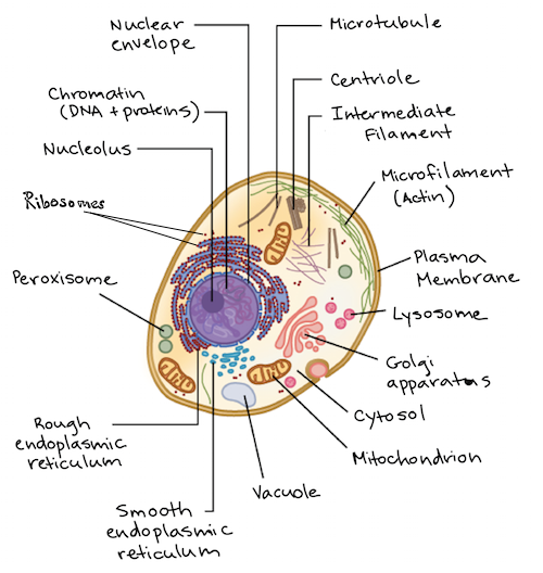
Image modified from OpenStax Biology.
Diagram of a typical plant cell:
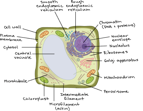
Image modified from OpenStax Biology.
Plasma membrane and cytoplasm
Introduction
What’s a cell? Well, on some level, it's a bag of goo. The plasma membrane—the outer boundary of the cell—is the bag, and the cytoplasm is the goo.
Of course, a cell is ever so much more than just a bag of goo. It's a complex, highly organized unit, the basic building block of all living things. And the plasma membrane and cytoplasm are actually pretty sophisticated.
The membrane is a delicate, two-layered structure of lipids and proteins, and it controls what can enter and exit the cell. Similarly, the cytoplasm of a eukaryotic cell consists not only of cytosol—a gel-like substance made up of water, ions, and macromolecules—but also of organelles and the structural proteins that make up the cytoskeleton, or "skeleton of the cell."
In this article, we’ll take a closer look at the plasma membrane and cytoplasm.
The plasma membrane
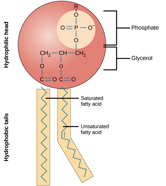
Both prokaryotic and eukaryotic cells have a plasma membrane, a double layer of lipids that separates the cell interior from the outside environment. This double layer consists largely of specialized lipids called phospholipids.
A phospholipid is made up of a hydrophilic, water-loving, phosphate head, along with two hydrophobic, water-fearing, fatty acid tails. Phospholipids spontaneously arrange themselves in a double-layered structure with their hydrophobic tails pointing inward and their hydrophilic heads facing outward. This energetically favorable two-layer structure, called a phospholipid bilayer, is found in many biological membranes.
As shown below, proteins are also an important component of the plasma membrane. Some of them pass all the way through the membrane, serving as channels or signal receptors, while others are just attached at the edge. Different types of lipids, such as cholesterol, may also be found in the cell membrane and affect its fluidity.
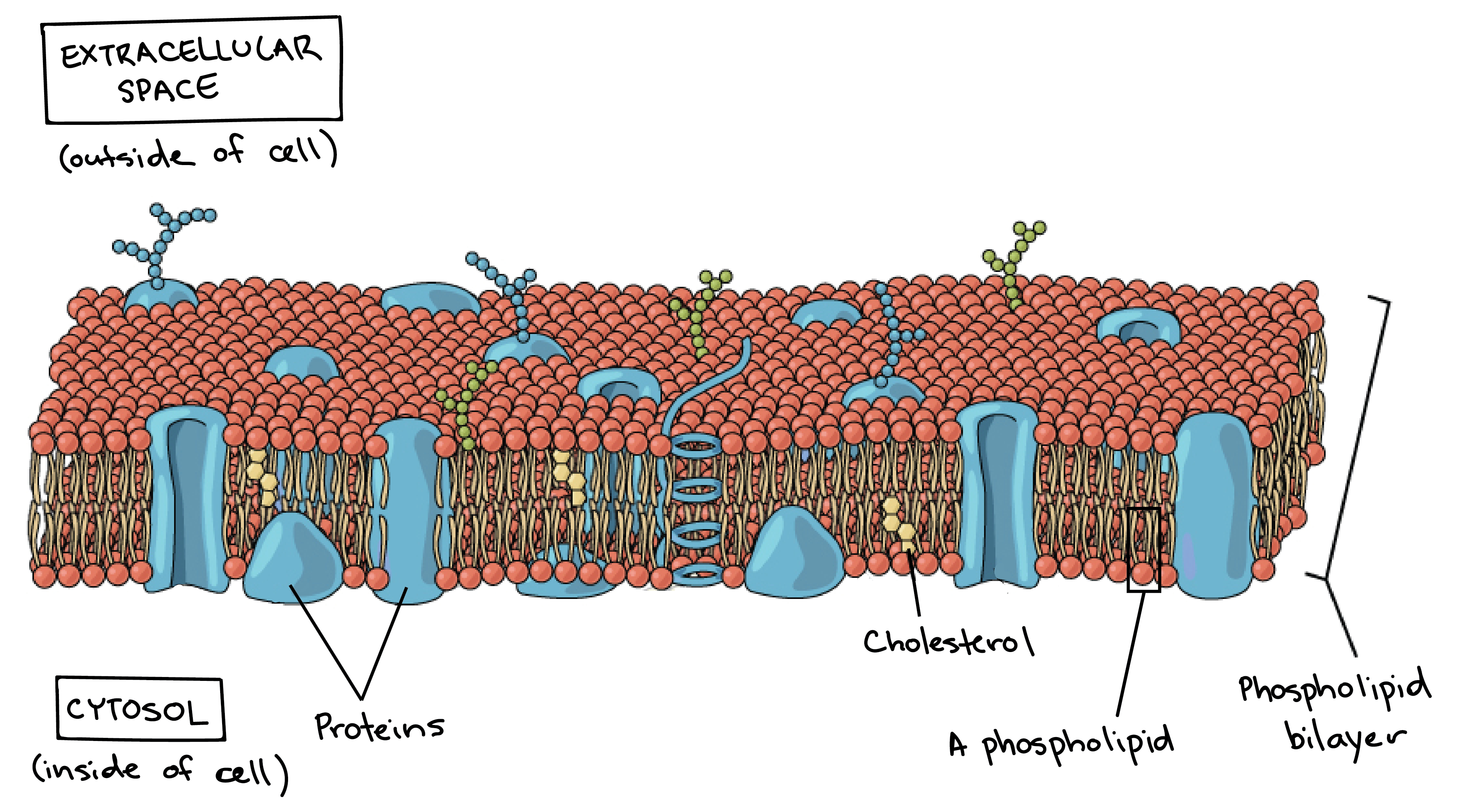
Image credit: modified from OpenStax Biology
The plasma membrane is the border between the interior and exterior of a cell. As such, it controls passage of various molecules—including sugars, amino acids, ions, and water—into and out of the cell. How easily these molecules can cross the membrane depends on their size and polarity. Some small, nonpolar molecules, such as oxygen, can pass directly through the phospholipid portion of the membrane. Larger and more polar, hydrophilic, molecules, such as amino acids, must instead cross the membrane by way of protein channels, a process that is often regulated by the cell. You can learn more about cellular transport in the membranes and transport section.
The surface area of the plasma membrane limits the exchange of materials between a cell and its environment. Some cells are specialized in the exchange of wastes or nutrients and have modifications to increase the area of the plasma membrane. For instance, the membranes of some nutrient-absorbing cells are folded into fingerlike projections called microvilli, singular, microvillus. Cells with microvilli cover the inside surface of the small intestine, the organ that absorbs nutrients from digested food. The microvilli help intestinal cells maximize their absorption of nutrients from food by increasing plasma membrane surface area.
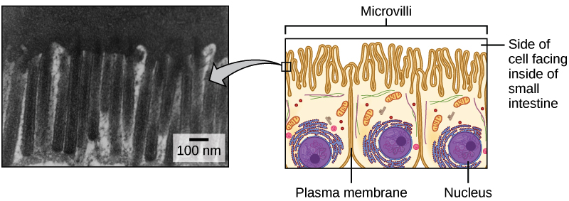
Image credit: OpenStax Biology. Micrograph is a modification of work by Louisa Howard.
The cytoplasm
The part of the cell referred to as cytoplasm is slightly different in eukaryotes and prokaryotes. In eukaryotic cells, which have a nucleus, the cytoplasm is everything between the plasma membrane and the nuclear envelope. In prokaryotes, which lack a nucleus, cytoplasm simply means everything found inside the plasma membrane.
One major component of the cytoplasm in both prokaryotes and eukaryotes is the gel-like cytosol, a water-based solution that contains ions, small molecules, and macromolecules. In eukaryotes, the cytoplasm also includes membrane-bound organelles, which are suspended in the cytosol. The cytoskeleton, a network of fibers that supports the cell and gives it shape, is also part of the cytoplasm and helps to organize cellular components.
Even though the cytosol is mostly water, it has a semi-solid, Jello-like consistency because of the many proteins suspended in it. The cytosol contains a rich broth of macromolecules and smaller organic molecules, including glucose and other simple sugars, polysaccharides, amino acids, nucleic acids, and fatty acids. Ions of sodium, potassium, calcium, and other elements are also found in the cytosol. Many metabolic reactions, including protein synthesis, take place in this part of the cell.
Nucleus and ribosomes
Introduction
Suppose that you have a very precious piece of information. Let’s imagine that this piece of information is a blueprint. In fact, it’s not just a blueprint for a house, or a car, or even a top-secret fighter jet. It’s a blueprint for an entire organism – you – and it not only specifies how to put you together, but also provides the information that enables every cell in your body to keep functioning from moment to moment.
Sounds important, right? You’d probably want to keep information this valuable in a secure spot, perhaps in a protected vault where you can keep an eye on it. In fact, that’s exactly what eukaryotic cells do with their genetic material, placing it in a membrane-enclosed repository called the nucleus.
Eukaryotic DNA never leaves the nucleus; instead, it’s transcribed (copied) into RNA molecules, which may then travel out of the nucleus. In the cytosol, some RNAs associate with structures called ribosomes, where they direct synthesis of proteins. (Other RNAs play functional roles in the cell, serving as structural components of the ribosome or regulating activity of genes.) Here, we’ll look in a little more detail at the structure of the nucleus and ribosomes.
The nucleus
The nucleus (plural, nuclei) houses the cell’s genetic material, or DNA, and is also the site of synthesis for ribosomes, the cellular machines that assemble proteins. Inside the nucleus, chromatin (DNA wrapped around proteins, described further below) is stored in a gel-like substance called nucleoplasm.
Enclosing the nucleoplasm is the nuclear envelope, which is made up of two layers of membrane: an outer membrane and an inner membrane. Each of these membranes contains two layers of phospholipids, arranged with their tails pointing inward (forming a phospholipid bilayer). There’s a thin space between the two layers of the nuclear envelope, and this space is directly connected to the interior of another membranous organelle, the endoplasmic reticulum.
Nuclear pores, small channels that span the nuclear envelope, let substances enter and exit the nucleus. Each pore is lined by a set of proteins, called the nuclear pore complex, that control what molecules can go in or out.
If you look at a microscope image of the nucleus, you may notice – depending on the type of stain used to visualize the cell – that there’s a dark spot inside it. This darkly staining region is called the nucleolus, and it’s the site in which new ribosomes are assembled.
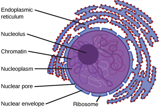
Image credit: OpenStax Biology.
How do you make a ribosome? Some chromosomes have sections of DNA that encode ribosomal RNA, a type of structural RNA that combines with proteins to make the ribosome. In the nucleolus, new ribosomal RNA combines with proteins to form the subunits of the ribosome. The newly made subunits are transported out through the nuclear pores to the cytoplasm, where they can do their job.
Some cell types have more than one nucleolus inside the nucleus. For instance, some mouse cells have up to \[6\] nucleoli\[^1\]. Prokaryotes, which do not have a nucleus, don't have nucleoli and build their ribosomes in the cytosol.
Chromosomes and DNA
Now that we have a sense of the structure of the nucleus, let’s have a closer look at the genetic information stored inside it: the DNA. Most of an organism’s DNA is organized into one or more chromosomes, each of which is a very long string or loop of DNA. A single chromosome can carry many different genes.
In prokaryotes, DNA is typically organized into a single circular chromosome (a loop). In eukaryotes, on the other hand, chromosomes are linear structures (strings). Every eukaryotic species has a specific number of chromosomes in the nuclei of its body’s cells. For example, a typical human body cell would have \[46\] chromosomes, while a comparable fruit fly cell would have \[8\].
Chromosomes are only visible as distinct structures when the cell is getting ready to divide. When the cell is in the growth and maintenance phases of its life cycle, the chromosomes instead resemble an unwound, jumbled bunch of threads. In this form, the DNA is accessible to the enzymes that transcribe it into RNA, allowing the genetic information to be put to use (expressed).
In both their loose and compact forms, the DNA strands of chromosomes are bound to structural proteins, including a family of proteins called histones (see picture below). These DNA-associated proteins organize the DNA and help it fit into the nucleus, and they also play a role in determining which genes are active or inactive. The complex formed by DNA and its supporting structural proteins is known as chromatin. You can learn more about DNA, chromatin, and chromosomes in the DNA and chromosomes article.
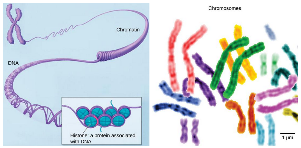
Image credit: OpenStax Biology. Image at right is a modification of work by the NIH; scale-bar data from Matt Russell.
To give you a sense of just how important DNA packing is, consider that the DNA in a typical human cell would be about \[2\] meters long if it were extended in a straight line. All \[2\] meters of that DNA are squeezed into a tiny nucleus with a diameter of just \[0.006\] mm. That's a feat "geometrically equivalent to packing \[40\] km (\[24\] miles) of extremely fine thread into a tennis ball" \[^4\]!
Ribosomes
As mentioned above, ribosomes are the molecular machines responsible for protein synthesis. A ribosome is made out of RNA and proteins, and each ribosome consists of two separate RNA-protein complexes, known as the small and large subunits. The large subunit sits on top of the small subunit, with an RNA template sandwiched between the two. (A ribosome looks a little like a hamburger with a puffy bun on top, an RNA “patty” threading through it.)
In eukaryotes, ribosomes get their orders for protein synthesis from the nucleus, where portions of DNA (genes) are transcribed to make messenger RNAs (mRNAs). An mRNA travels to the ribosome, which uses the information it contains to build a protein with a specific amino acid sequence. This process is called translation. Prokaryotes lack a nucleus, so their mRNAs are transcribed in the cytoplasm and can be translated by ribosomes immediately.
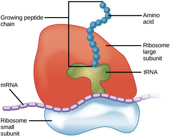
Image credit: OpenStax Biology.
Eukaryotic ribosomes may be either free, meaning that they are floating around in the cytoplasm, or bound, meaning that they are attached to the endoplasmic reticulum or the outside of the nuclear envelope. (In the first diagram in this article, the red dots represent bound ribosomes; endoplasmic reticulum with bound ribosomes is known as rough endoplasmic reticulum.)
Because protein synthesis is an essential function of all cells, ribosomes are found in practically every cell type of multicellular organisms, as well as in prokaryotes such as bacteria. However, eukaryotic cells that specialize in producing proteins have particularly large numbers of ribosomes. For example, the pancreas is responsible for producing and secreting large amounts of digestive enzymes, so the pancreatic cells that make these enzymes have an unusually high number of ribosomes.
Final fun fact: in a testament to the importance of the ribosome, the 2009 Nobel Prize in Chemistry was awarded to three researchers who mapped its structure and movements down to the level of individual atoms using a technique called X-ray crystallography\[^5\].
Prokaryotes and eukaryotes review
Key terms
| Term | Meaning |
|---|---|
| Endosymbiotic theory | Theory proposing that eukaryotic cells formed from a symbiotic relationship among prokaryotic cells |
How do prokaryotic and eukaryotic cells differ?
| Prokaryotes | Eukaryotes | ||
|---|---|---|---|
| Genetic information | DNA is circular, usually free-floating in cytoplasm | DNA is linear, found in nucleus | |
| Organelles | No nucleus or membrane-bound organelles | Has nucleus and membrane-bound organelles (ie: mitochondria, chloroplasts, Golgi body, ER) | |
| Size | Small (1-5 micrometers) | Larger (10-100 micrometers) | |
| Organisms | Bacteria/archaea | Animals, plants, fungi, protists | |
| Cell structure | Always unicellular | Can be unicellular or multicellular |
What is the endosymbiotic theory?
One theory that may explain how eukaryotes became so complex is the endosymbiotic theory.
This theory proposes that organelles like mitochondria and chloroplasts were once free-living prokaryotic cells that began to live within a larger host cell. Over a long time, the prokaryotes and their hosts evolved together until one could not function without the other.
Common mistakes and misconceptions
Eukaryotes can be unicellular. Many people think that eukaryotes are all multicellular, but this is not the case. While prokaryotes are always unicellular organisms, eukaryotes can be either unicellular or multicellular. For example, most protists are single-celled eukaryotes!
Even though prokaryotes do not have a nucleus, they DO contain genetic information. Prokaryotes generally have single circular chromosomes where they store their genetic information.