Organelles in eukaryotic cells
Endoplasmic reticulum and Golgi bodies
Endomembrane system
The endomembrane system
Introduction
Let’s imagine you are a pancreatic cell. Your job is to secrete digestive enzymes, which travel into the small intestine and help break down nutrients from food. In order to carry out this job, you somehow have to get those enzymes shipped from their site of synthesis—inside the cell—to their place of action—outside the cell.
How are you going to make this happen? After a moment of panic in which you consider calling the postal service, you relax, having remembered: I have an endomembrane system!
What is the endomembrane system?
The endomembrane system (endo- = “within”) is a group of membranes and organelles in eukaryotic cells that works together to modify, package, and transport lipids and proteins. It includes a variety of organelles, such as the nuclear envelope and lysosomes, which you may already know, and the endoplasmic reticulum and Golgi apparatus, which we will cover shortly.
Although it's not technically inside the cell, the plasma membrane is also part of the endomembrane system. As we'll see, the plasma membrane interacts with the other endomembrane organelles, and it's the site where secreted proteins (like the pancreatic enzymes in the intro) are exported. Important note: the endomembrane system does not include mitochondria, chloroplasts, or peroxisomes.
Let's take a closer look at the different parts of the endomembrane system and how they function in the shipping of proteins and lipids.
The endoplasmic reticulum
The endoplasmic reticulum (ER) plays a key role in the modification of proteins and the synthesis of lipids. It consists of a network of membranous tubules and flattened sacs. The discs and tubules of the ER are hollow, and the space inside is called the lumen.
Rough ER
The rough endoplasmic reticulum (rough ER) gets its name from the bumpy ribosomes attached to its cytoplasmic surface. As these ribosomes make proteins, they feed the newly forming protein chains into the lumen. Some are transferred fully into the ER and float inside, while others are anchored in the membrane.
Inside the ER, the proteins fold and undergo modifications, such as the addition of carbohydrate side chains. These modified proteins will be incorporated into cellular membranes—the membrane of the ER or those of other organelles—or secreted from the cell.
If the modified proteins are not destined to stay in the ER, they will be packaged into vesicles, or small spheres of membrane that are used for transport, and shipped to the Golgi apparatus. The rough ER also makes phospholipids for other cellular membranes, which are transported when the vesicle forms.
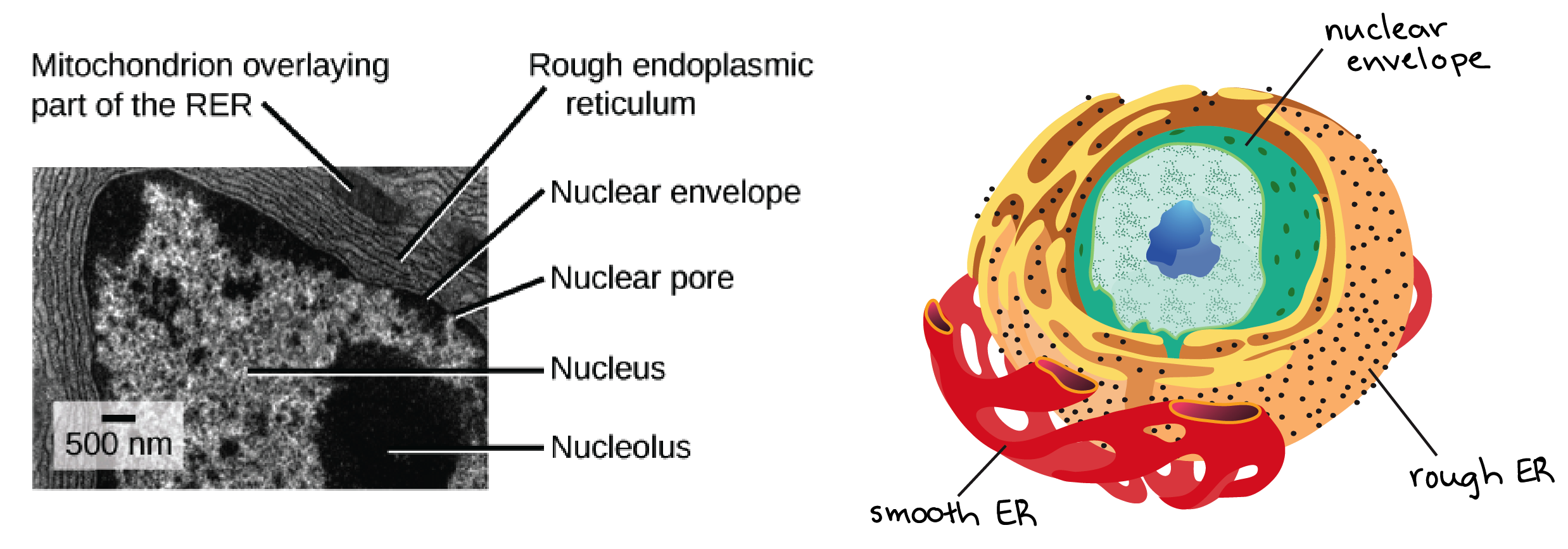
_Image credit: left, "The endomembrane system and proteins: Figure 2" by OpenStax College, Biology (CC BY 3.0), modification of work by Lousia Howard; right, modification of "Animal cell structure" by Mariana Ruiz, public domain_
Since the rough ER helps modify proteins that will be secreted from the cell, cells whose job is to secrete large amounts of enzymes or other proteins, such as liver cells, have lots of rough ER.
Smooth ER
The smooth endoplasmic reticulum (smooth ER) is continuous with the rough ER but has few or no ribosomes on its cytoplasmic surface. Functions of the smooth ER include:
- Synthesis of carbohydrates, lipids, and steroid hormones
- Detoxification of medications and poisons
- Storage of calcium ions
In muscle cells, a special type of smooth ER called the sarcoplasmic reticulum is responsible for storage of calcium ions which are needed to trigger the coordinated contractions of muscle fibers.
There are also tiny "smooth" patches of ER found within the rough ER. These patches serve as exit sites for vesicles budding off from the rough ER and are called transitional ER\[^1\].
The Golgi apparatus
When vesicles bud off from the ER, where do they go? Before reaching their final destination, the lipids and proteins in the transport vesicles need to be sorted, packaged, and tagged so that they wind up in the right place. This sorting, tagging, packaging, and distribution takes place in the Golgi apparatus (Golgi body), an organelle made up of flattened discs of membrane.
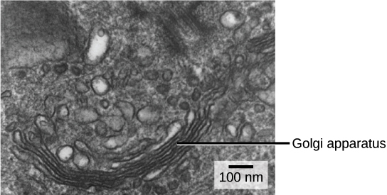
_Image credit: "The endomembrane system and proteins: Figure 3" by OpenStax College, Biology (CC BY 3.0), modification of work by Lousia Howard_
The receiving side of the Golgi apparatus is called the cis face and the opposite side is called the trans face. Transport vesicles from the ER travel to the cis face, fuse with it, and empty their contents into the lumen of the Golgi apparatus.
As proteins and lipids travel through the Golgi, they undergo further modifications. Short chains of sugar molecules might be added or removed, or phosphate groups attached as tags. Carbohydrate processing is shown in the diagram as the gain and loss of branches on the purple carbohydrate group attached to the protein.
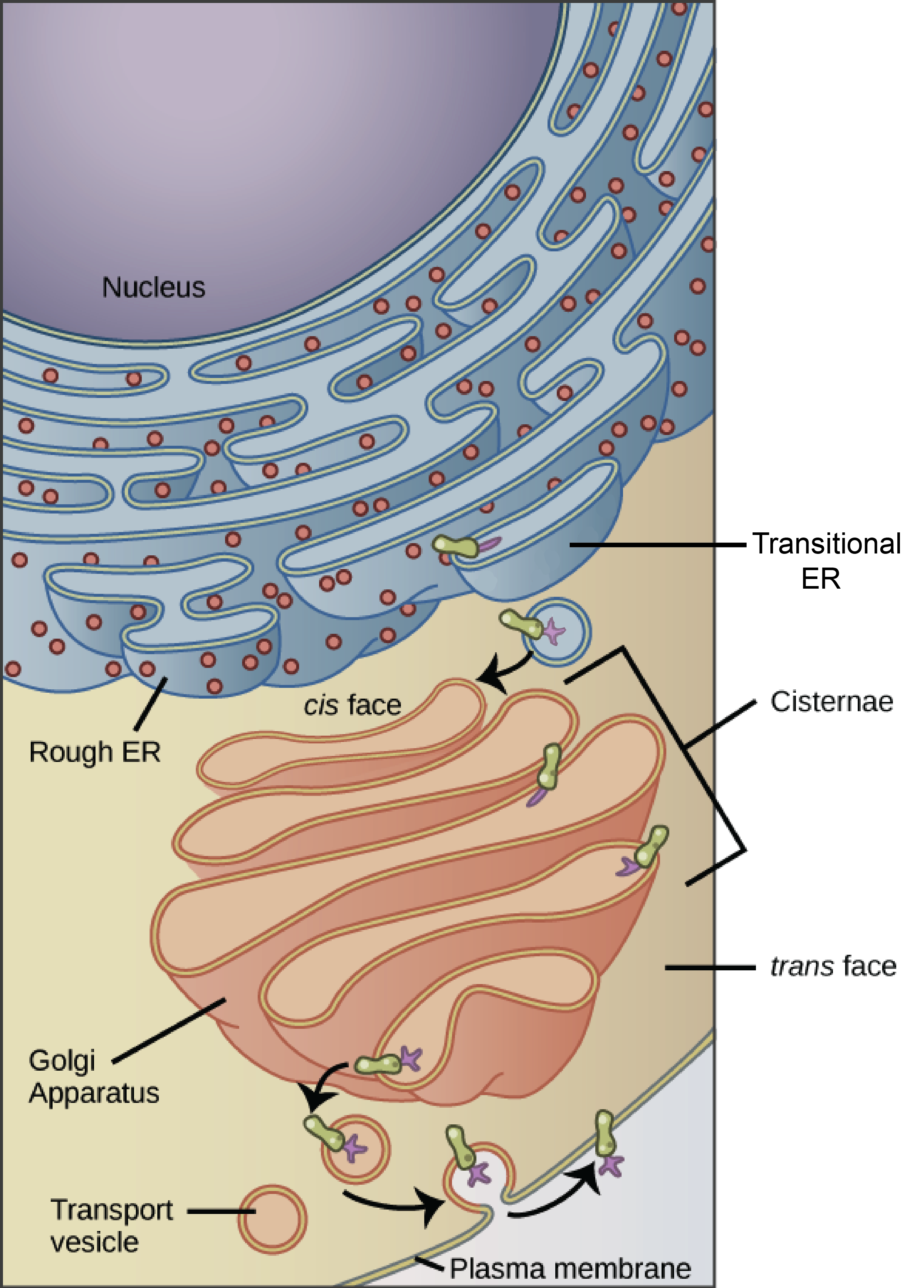
_Image modified from "The endomembrane system and proteins: Figure 1" by OpenStax College, Biology (CC BY 3.0), modification of work by Magnus Manske_
Finally, the modified proteins are sorted (based on markers such as amino acid sequences and chemical tags) and packaged into vesicles that bud from the trans face of the Golgi. Some of these vesicles deliver their contents to other parts of the cell where they will be used, such as the lysosome or vacuole. Others fuse with the plasma membrane, delivering membrane-anchored proteins that function there and releasing secreted proteins outside the cell.
Cells that secrete many proteins—such as salivary gland cells that secrete digestive enzymes, or cells of the immune system that secrete antibodies—have many Golgi stacks. In plant cells, the Golgi apparatus also makes polysaccharides (long-chain carbohydrates), some of which are incorporated into the cell wall.
Lysosomes
The lysosome is an organelle that contains digestive enzymes and acts as the organelle-recycling facility of an animal cell. It breaks down old and unnecessary structures so their molecules can be reused. Lysosomes are part of the endomembrane system, and some vesicles that leave the Golgi are bound for the lysosome.
Lysosomes can also digest foreign particles that are brought into the cell from outside. As an example, let's consider a class of white blood cells called macrophages, which are part of the human immune system. In a process known as phagocytosis, a section of the macrophage’s plasma membrane invaginates—folds inward—to engulf a pathogen, as shown below.
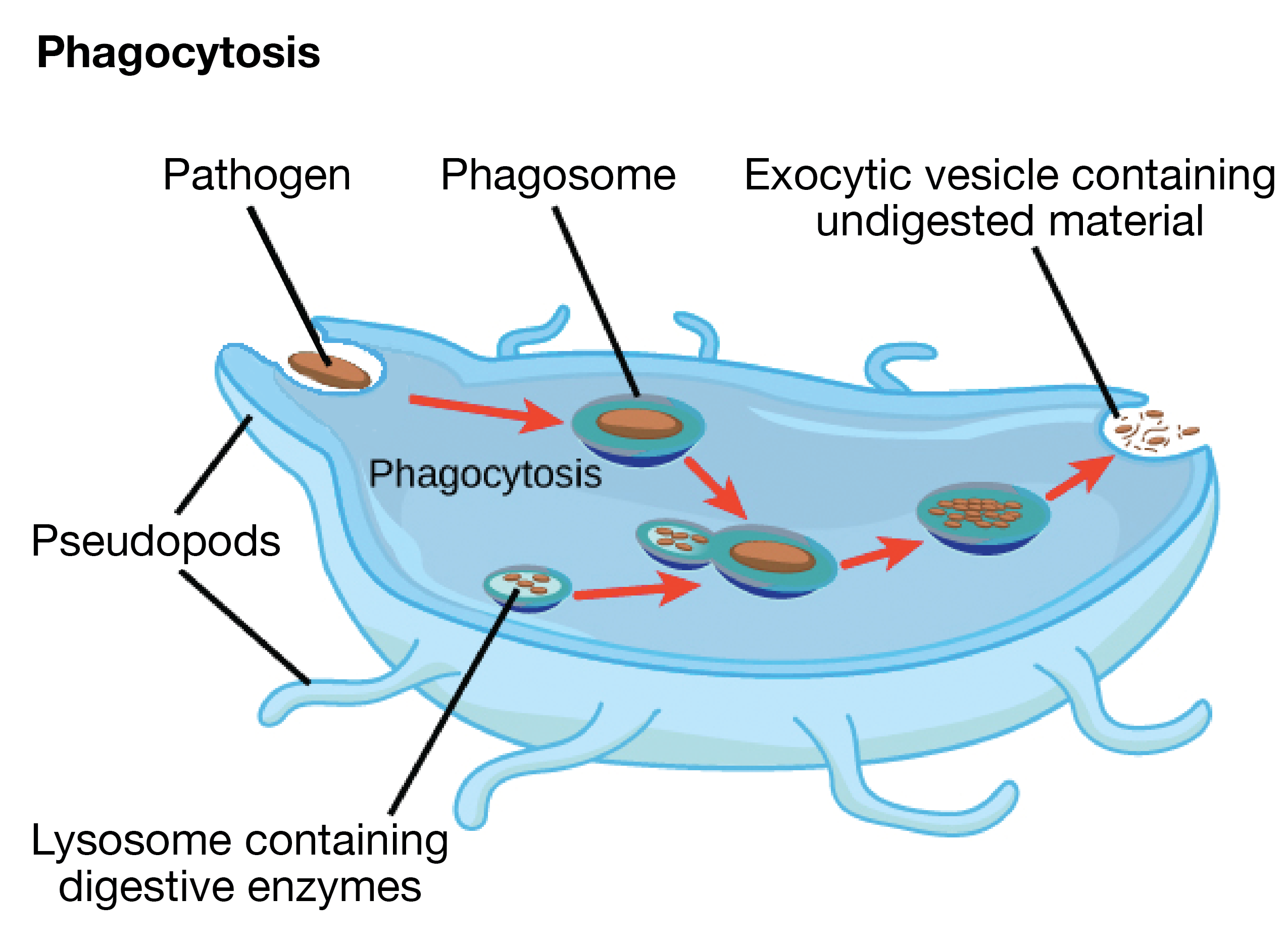
_Image credit: modified from "The endomembrane system and proteins: Figure 4" by OpenStax College, Biology (CC BY 3.0)_
The invaginated section, with the pathogen inside, pinches off from the plasma membrane to form a structure called a phagosome. The phagosome then fuses with a lysosome, forming a combined compartment where digestive enzymes destroy the pathogen.
Vacuoles
Plants cells are unique because they have a lysosome-like organelle called the vacuole. The large central vacuole stores water and wastes, isolates hazardous materials, and has enzymes that can break down macromolecules and cellular components, like those of a lysosome.\[^3\] Plant vacuoles also function in water balance and may be used to store compounds such as toxins and pigments (colored particles).\[^4\]
Lysosomes vs. peroxisomes
One point that can be confusing is the difference between lysosomes and peroxisomes. Both types of organelles are involved in breaking down molecules and neutralizing hazards to the cell. Also, both usually show up as small, round blobs in diagrams.
However, the peroxisome is a different organelle with its own unique properties and role in the cell. It houses enzymes involved in oxidation reactions, which produce hydrogen peroxide (\[\text H_2 \text O_2\]) as a by-product. The enzymes break down fatty acids and amino acids, and they also detoxify some substances that enter the body. For example, alcohol is detoxified by peroxisomes found in liver cells.
Importantly, peroxisomes—unlike lysosomes—are not part of the endomembrane system. That means they don't receive vesicles from the Golgi apparatus. You can learn more about how proteins are shipped to the peroxisome in the article on protein targeting.
Mitochondria
Mitochondria and chloroplasts
Key points:
Mitochondria are the "powerhouses" of the cell, breaking down fuel molecules and capturing energy in cellular respiration.
Chloroplasts are found in plants and algae. They're responsible for capturing light energy to make sugars in photosynthesis.
Mitochondria and chloroplasts likely began as bacteria that were engulfed by larger cells (the endosymbiont theory).
Introduction
You may know that your body is made up of cells (trillions and trillions of them). You may also know that the reason you need to eat food—such as veggies—is so that you have the energy to do things like play sports, study, walk, and even breathe.
But what exactly happens in your body to turn the food energy stored in broccoli into a form that your body can use? And how does energy end up stored in the broccoli to begin with, anyway?
The answers to these questions have a lot to do with two important organelles: mitochondria and chloroplasts.
Chloroplasts are organelles found in the broccoli's cells, along with those of other plants and algae. They capture light energy and store it as fuel molecules in the plant's tissues.
Mitochondria are found inside of your cells, along with the cells of plants. They convert the energy stored in molecules from the broccoli (or other fuel molecules) into a form the cell can use.
Let's take a closer look at these two very important organelles.
Chloroplasts
Chloroplasts are found only in plants and photosynthetic algae. (Humans and other animals do not have chloroplasts.) The chloroplast's job is to carry out a process called photosynthesis.
In photosynthesis, light energy is collected and used to build sugars from carbon dioxide. The sugars produced in photosynthesis may be used by the plant cell, or may be consumed by animals that eat the plant, such as humans. The energy contained in these sugars is harvested through a process called cellular respiration, which happens in the mitochondria of both plant and animal cells.
Chloroplasts are disc-shaped organelles found in the cytosol of a cell. They have outer and inner membranes with an intermembrane space between them. If you passed through the two layers of membrane and reached the space in the center, you’d find that it contained membrane discs known as thylakoids, arranged in interconnected stacks called grana (singular, granum).
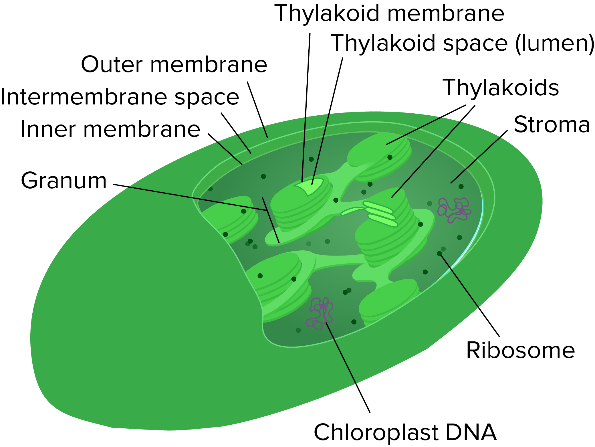
_Image modified from "Chloroplast mini," by Kelvin Ma (CC BY 3.0)._
The membrane of a thylakoid disc contains light-harvesting complexes that include chlorophyll, a pigment that gives plants their green color. Thylakoid discs are hollow, and the space inside a disc is called the thylakoid space or lumen, while the fluid surrounding the thylakoids is called the stroma.
You can learn more about chloroplasts, chlorophyll, and photosynthesis in the photosynthesis topic section.
Mitochondria
Mitochondria (singular, mitochondrion) are often called the powerhouses or energy factories of the cell. Their job is to make a steady supply of adenosine triphosphate (ATP), the cell’s main energy-carrying molecule. The process of making ATP using chemical energy from fuels such as sugars is called cellular respiration, and many of its steps happen inside the mitochondria.
The mitochondria are suspended in the jelly-like cytosol of the cell. They are oval-shaped and have two membranes: an outer one, surrounding the whole organelle, and an inner one, with many inward protrusions called cristae that increase surface area.
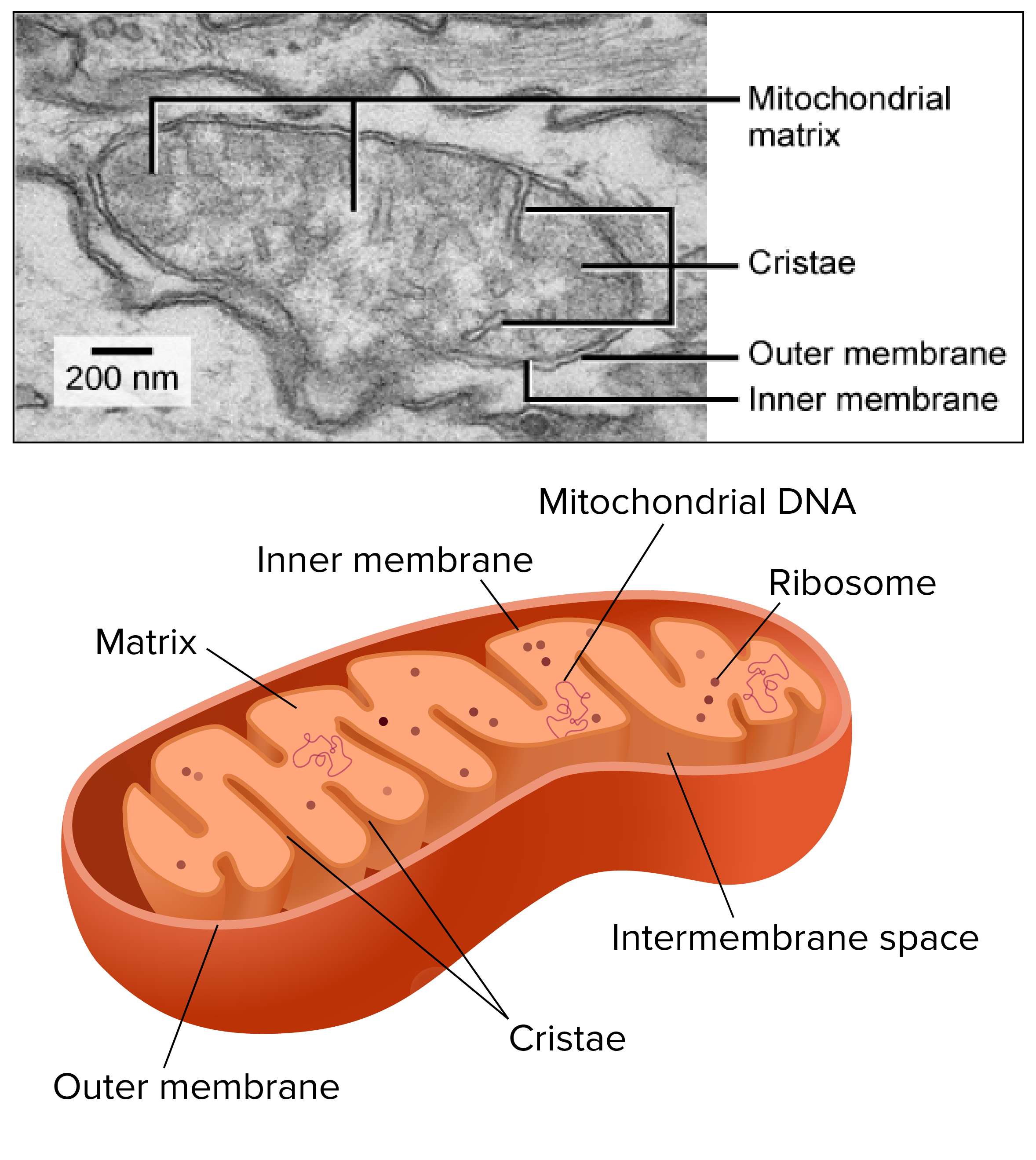
_Image credits: upper image, "Eukaryotic cells: Figure 7," by OpenStax College, Biology (CC BY 3.0). Modification of work by Matthew Britton; scale-bar data from Matt Russell. Lower image: modification of "Mitochondrion mini," by Kelvin Ma (public domain)._
Cristae were once thought to be broad, wavy folds, but as Sal discusses in his mitochondria video, they're now understood to be more like long caverns.\[^1\] Here is a 3D reconstruction of a slice cut from a mitochondrion:
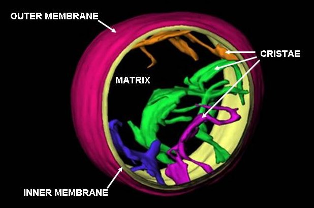
Image credit: "MitochondrionCAM," by Carmann (public domain).\[^2\]
The space between the membranes is called the intermembrane space, and the compartment enclosed by the inner membrane is called the mitochondrial matrix. The matrix contains mitochondrial DNA and ribosomes. We'll talk shortly about why mitochondria (and chloroplasts) have their own DNA and ribosomes.
The multi-compartment structure of the mitochondrion may seem complicated to us. That's true, but it turns out to be very useful for cellular respiration, allowing reactions to be kept separate and different concentrations of molecules to be maintained in different "rooms."
Although mitochondria are found in most human cell types (as well as most cell types in other animals and plants), their numbers vary depending on the role of the cell and its energy needs. For instance, muscle cells typically have high energy needs and large numbers of mitochondria, while red blood cells, which are highly specialized for oxygen transport, have no mitochondria at all.\[^3\]
Where did these organelles come from?
Both mitochondria and chloroplasts contain their own DNA and ribosomes. Why would these organelles need DNA and ribosomes, when there is DNA in the nucleus and ribosomes in the cytosol?
Strong evidence points to endosymbiosis as the answer to the puzzle. Symbiosis is a relationship in which organisms from two separate species live in a close, dependent relationship. Endosymbiosis (endo- = “within”) is a specific type of symbiosis where one organism lives inside the other.
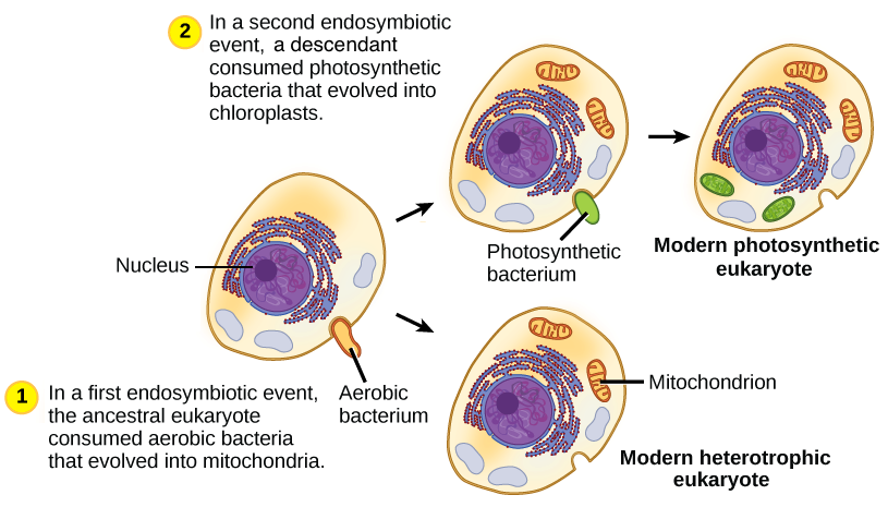
_Image modified from "Eukaryotic origins: Figure 4," by OpenStax College, Biology, (CC BY 4.0)._
Bacteria, mitochondria, and chloroplasts are similar in size. Bacteria also have DNA and ribosomes similar to those of mitochondria and chloroplasts.\[^4\] Based on this and other evidence, scientists think host cells and bacteria formed endosymbiotic relationships long ago, when individual host cells took in aerobic (oxygen-using) and photosynthetic bacteria but did not destroy them. Through millions of years of evolution, the aerobic bacteria became mitochondria and the photosynthetic bacteria became chloroplasts.